For additional assistance, please contact support@metavilabs.com.
Gallery
Note: no labels are Dyes are used in these demonstrations. All colors are added by FastTrack AI based on cell characteristics in bright-field or phase-contrast.
Demonstration of FastTrack AI measuring T cell killing efficiency on the ibidi adhesion pad grid slide. The source movie is phase contrast with no dyes or labels. The AI system tags the imune cells with a green mark and the letter T. The AI system tags the cancer cells with a blue mark and the letter P (pathogen). The AI system tags dead cell matter with red marks. Pad locations with an initial living cancer cell are marked with a blue square. Pad locations which loose the living cell are marked with a Red square. The blue to red transition marks a living to dead cell transition.
Demonstration of FastTrack AI tagging T cells with a green mark, tagging cancer cells with a red mark; bright-field, co-culture of CAR T cells and Cancer Cells. No labels or dyes are used.
Demonstration of FastTrack AI tagging apoptotic bodies with a green mark; bright-field, co-culture of CAR T cells and Cancer Cells. Please watch to the end, the apoptotic bodies (due to CAR T cell killing) appear near the end of the time-lapse. No labels or dyes are used.
Demonstration of FastTrack AI tagging all cells; bright-field, co-culture of CAR T cells and Cancer Cells.
Demonstration of FastTrack AI tagging and tracking cells in an ibidi chemotaxis chamber; phase contrast, cancer cells.
Demonstration of FastTrack AI tagging cells as they change shape; phase contrast, cancer cells.
Demonstration of FastTrack AI tagging cells shape changes on a collagen coated surface; phase contrast, fibroblasts cells.
Demonstration of FastTrack AI tagging nuetrophils on 2D glass in bright-field.
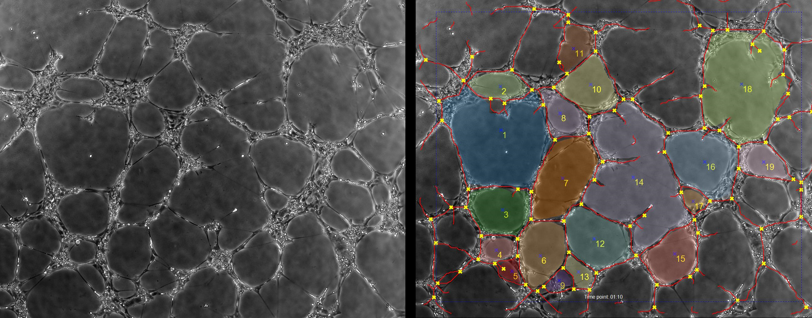
Demonstration of Angiogenesis-Tube-formation analysis of phase contrast .